Debridement, Irrigation, and Decompression of the Bone for Chronic Osteomyelitis of the Distal Tibia and Active Sinuses
Samhita Kadiyala, BA1; Donald L. Fejfar, MD1; Nikhil Gattu, MD1; Emily S. Powis, BS1; Elizabeth Rich, MD2; Hizkyas Kassaye, MD3; Wesley Kalomba, MD4; Ridwan M. Saeed, MD5; Bitiel Banda, MD6; Pierre M. Woolley, MD7; Kiran J. Agarwal-Harding, MD, MPH1,8
1Harvard Global Orthopaedics Collaborative, Boston, MA
2Walter Reed National Military Medical Center, Bethesda, MD
3Hawassa University Comprehensive Specialized Hospital, Hawassa, Ethiopia
4Lilongwe Institute of Orthopaedics and Neurosurgery, Lilongwe, Malawi
5Muhimbili Orthopaedic Institute, Dar es Salaam, Tanzania
6Mzuzu Central Hospital, Mzuzu, Malawi
7Hôpital Universitaire La Paix, Port au Prince, Haiti
8Beth Israel Deaconess Medical Center, Boston, MA
Chronic osteomyelitis (COM) is a debilitating bone infection that presents a significant health burden in low- and middle-income countries. In children, it frequently results from delayed treatment of acute hematogenous infection. This case involves a 13-year-old male presenting with over one month of leg pain, difficulty walking, and three active draining sinus tracts along the medial distal tibia. Preoperative radiographs revealed a sequestrum involving nearly half of the distal tibia with an incomplete involucrum.
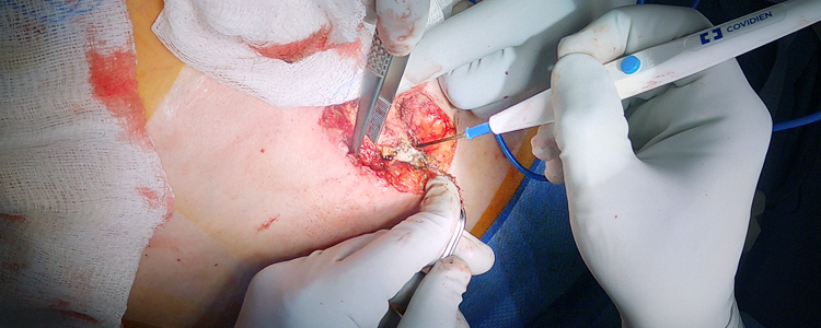
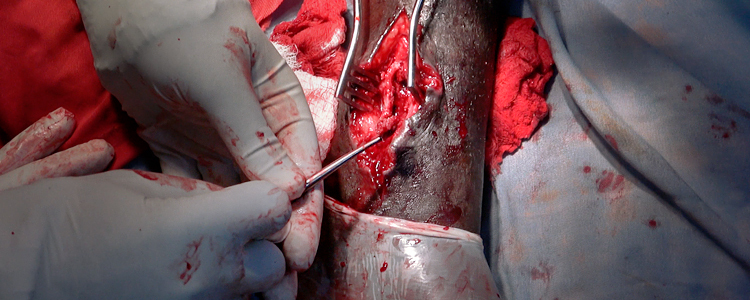
Surgical management focused on thorough debridement and decompression. The procedure involved making an incision to connect the sinus tracts, performing a sequestrectomy to clear the medullary canal, and using a drill to decompress a Brodie’s abscess. Debridement was continued until healthy bleeding bone was observed. Following saline and iodine irrigation, a gentamicin solution was applied as a local antibiotic adjunct. This case highlights the importance of balancing radical debridement with the preservation of structural stability in resource-limited settings. Postoperatively, the patient received broad-spectrum antibiotics and was maintained on partial weight-bearing to allow the involucrum to mature.