PUBLISHED: Superficial Femoral to Below-the-Knee Popliteal Artery Bypass for Popliteal Artery Aneurysm Containing Mural Thrombus

Superficial Femoral to Below-the-Knee Popliteal Artery Bypass for Popliteal Artery Aneurysm Containing Mural Thrombus
Brett J. Salomon, MD1,2; Tiffany R. Bellomo, MD1,2; Zachary M. Feldman, MD, MS1,2
1Massachusetts General Hospital
2Heart and Vascular Institute, Mass General Brigham

Popliteal artery aneurysms (PAAs) are the most common peripheral arterial aneurysms, with an incidence of approximately 10.4 cases per million per year and occur most frequently in men aged 60–70 years. Many lower extremity aneurysms are asymptomatic and are discovered incidentally on physical examination or imaging performed for unrelated conditions. However, symptoms resulting from compression of surrounding structures can occur and may be disabling, including deep vein thrombosis and neuropathies. The most serious complication is acute limb ischemia resulting from thrombosis or distal embolization. For this reason, the Society for Vascular Surgery (SVS) clinical practice guidelines recommend operative repair when an asymptomatic aneurysm reaches 20 mm in diameter or contains mural thrombus.

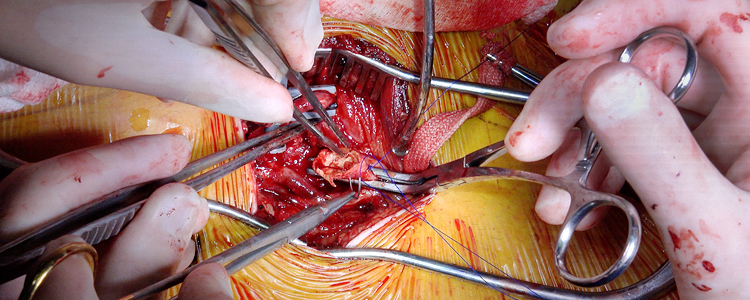
This article presents the case of a 67-year-old male with a 22-mm right PAA with significant mural thrombus. Given his right PAA was greater than 2 cm and had significant mural thrombus, he was evaluated for intervention. An intraoperative angiogram identified proximal and distal sites for bypass that were free of mural thrombus. Therefore, the patient underwent a superficial femoral to below-the-knee popliteal artery bypass using a great saphenous vein conduit through a medial incision in the supine position. The superficial femoral artery and below-the-knee popliteal artery were dissected free from the surrounding structures. The case required local thromboendarterectomy of the below-the-knee popliteal artery and tibioperoneal (TP) trunk, due to the presence of chronic thrombus at the endpoint that was not able to be visualized on preoperative angiography. A bypass was created between the two arteries, and the native artery was ligated, including the aneurysm.

The case highlights operative management of a PAA as well as the techniques involved in lower extremity bypass. Lower extremity bypass remains an important procedure for vascular and trauma surgeons who may need to perform this procedure for multiple pathologies.
PUBLISHED: Pediatric Supracondylar Humerus Fracture Repair with Mini-Open Assisted Reduction and Percutaneous Pinning

Pediatric Supracondylar Humerus Fracture Repair with Mini-Open Assisted Reduction and Percutaneous Pinning
Emily S. Powis, BS1; Donald L. Fejfar, MD1; Nikhil Gattu, MD1; Samhita Kadiyala, BA1; Elizabeth Rich, MD2; Wesley Kalomba, MD3; Hizkyas Kassaye, MD4; Ridwan M. Saeed, MD5; Bitiel Banda, MD6; Pierre M. Woolley, MD7; Kiran J. Agarwal-Harding, MD, MPH1,8
1Harvard Global Orthopaedics Collaborative, Boston, MA
2Walter Reed National Military Medical Center, Bethesda, MD
3Lilongwe Institute of Orthopaedics and Neurosurgery, Lilongwe, Malawi
4Hawassa University Comprehensive Specialized Hospital, Hawassa, Ethiopia
5Muhimbili Orthopaedic Institute, Dar es Salaam, Tanzania
6Mzuzu Central Hospital, Mzuzu, Malawi
7Hôpital Universitaire La Paix, Port au Prince, Haiti
8Beth Israel Deaconess Medical Center, Boston, MA

Supracondylar humerus (SCH) fractures are the most common elbow fractures in children and represent a significant source of morbidity in low- and middle-income countries (LMICs), where delayed presentation and limited surgical capacity frequently lead to poor outcomes. Closed reduction and percutaneous pinning (CRPP) is the gold-standard treatment for displaced SCH fractures and is particularly well-suited to resource-limited settings given its reliance on minimal, low-cost implants.

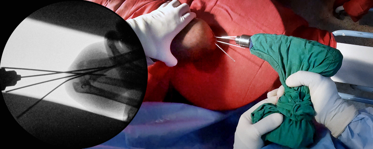
This case describes a 9-year-old male who presented to Mzuzu Central Hospital in Malawi with a completely displaced Gartland Type III SCH fracture following a fall. Surgical intervention was delayed three days due to unavailability of intraoperative fluoroscopic imaging. Neurovascular examination was intact on presentation. The procedure was performed under general anesthesia. Reduction was first attempted with longitudinal traction to unlock the distal fragment, followed by thumb pressure on the olecranon and elbow flexion. As the distal fragment remained incompletely reduced in the sagittal plane, a small posterior incision was made and the Kapandji maneuver was performed to achieve satisfactory reduction, confirmed fluoroscopically. Three smooth Kirschner wires (K-wires) were introduced in retrograde fashion from the lateral condyle under fluoroscopic guidance. One of the three K-wires was intramedullary and became loose prior to case conclusion, so it was removed before the remaining pins were cut and bent outside the skin, and a posterior back slab was applied with the elbow at 90 degrees of flexion. The remaining K-wires were removed at 4 weeks, and range-of-motion exercises were initiated thereafter.

This case demonstrates that percutaneous pinning, with mini-open assistance where closed reduction cannot be achieved, can be performed safely in resource-limited settings and illustrates the real-world intraoperative challenges, including limited C-arm resolution, use of non-orthopaedic drills with insufficient power, and bent K-wires, that afflict much of the developing world.